Skin Biopsy – Accurate Diagnosis for Healthy, Clear Skin
What Is a Skin Biopsy?
A skin biopsy involves removing a small sample of skin tissue for microscopic examination. This helps determine the exact cause of abnormal skin growths, rashes, infections, pigmentation changes, or suspected skin cancers.
It’s a quick, outpatient procedure performed under local anesthesia, ensuring minimal discomfort and zero downtime. The collected tissue sample is analyzed in a specialized laboratory to identify cellular changes or disease patterns.
Why Is a Skin Biopsy Needed?
Skin conditions often share similar surface symptoms — redness, itching, rashes, or lesions — making it difficult to pinpoint the root cause through visual examination alone. A biopsy provides a definitive diagnosis, helping dermatologists recommend the most suitable treatment plan.
Your dermatologist may suggest a skin biopsy if you have:
Unexplained rashes or chronic skin irritation
Non-healing wounds or sores
Suspicious moles or growths
Pigmentation disorders
Suspected skin infections
Signs of autoimmune or inflammatory skin diseases (like psoriasis, eczema, or lupus)
Potential skin cancer
By identifying the underlying issue at the cellular level, a skin biopsy ensures early intervention and effective management.
Types of Skin Biopsies
Depending on the skin concern, location, and depth of the affected area, your dermatologist will recommend one of the following types of biopsies:
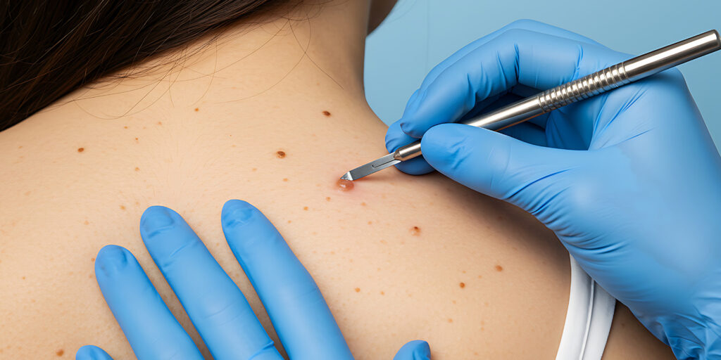
1. Punch Biopsy
A small, circular blade (punch tool) is used to remove a deeper sample of all skin layers — epidermis, dermis, and superficial fat.
Best for: Diagnosing inflammatory skin conditions, rashes, and deeper lesions.
Benefits:
Precise and minimally invasive
Quick healing
Accurate for microscopic diagnosis
2. Shave Biopsy
The top layers of the skin (epidermis and part of the dermis) are gently shaved using a sterile surgical blade.
Best for: Raised growths, warts, moles, or suspected basal cell carcinomas.
Benefits:
Simple, quick, and painless
No stitches required
Minimal scarring
3. Excisional Biopsy
The entire lesion or suspicious area is removed using a surgical scalpel, often with a small margin of surrounding healthy skin.
Best for: Suspected skin cancers, large moles, or cysts.
Benefits:
Complete lesion removal
Detailed examination of the entire sample
Reduces recurrence risk
4. Incisional Biopsy
Only a portion of a large lesion is removed for diagnostic purposes.
Best for: Extensive rashes or larger abnormal growths.
Benefits:
Ideal when the lesion covers a large area
Helps guide further treatment or surgery
The Skin Biopsy Procedure
Consultation & Evaluation:
The dermatologist evaluates your skin condition, discusses your medical history, and explains the procedure in detail.Local Anesthesia:
A mild anesthetic is applied to numb the biopsy site, ensuring a comfortable and pain-free experience.Tissue Sampling:
The dermatologist removes a small piece of skin using the appropriate biopsy technique.Wound Care:
The area is cleaned and dressed. In some cases, a few tiny stitches may be used, depending on the biopsy type.Laboratory Analysis:
The sample is sent to a dermatopathology lab for microscopic examination.Diagnosis & Treatment Planning:
Once the results are ready, your dermatologist explains the findings and recommends an individualized treatment plan.
Aftercare and Recovery
Proper post-biopsy care ensures fast healing and minimal scarring. Here’s what you can expect:
Keep the area clean and dry for 24–48 hours.
Change the dressing as instructed by your doctor.
Avoid scratching, rubbing, or wetting the wound.
Apply prescribed antibiotic ointments if recommended.
Stitches, if any, are usually removed in 7–10 days.
Healing time varies depending on the biopsy type and site but typically ranges from one to two weeks.